stratigraphical unit or a plate on a microscope
We now need to work out the entire length of the eyepiece graticule in the image above the eyepiece graticule reached 69 on the stage micrometer and so the whole length of the. The quarter wavelength retardation plate is a common optical.

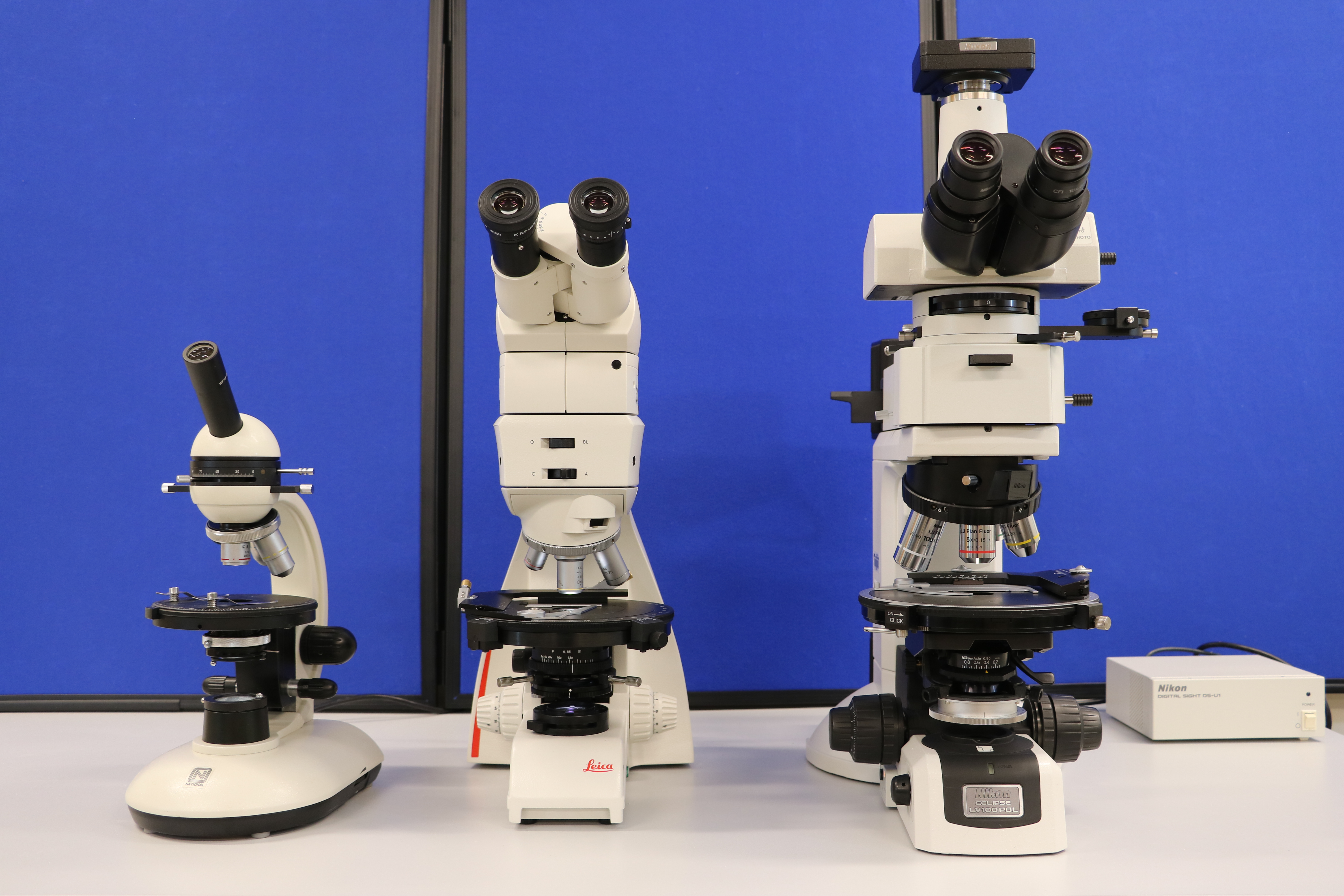
2 4 Parts Of The Petrographic Microscope Introduction To Petrology
Stratigraphy is a branch of geology to description of rock or interpretation geologic time scaleIt provides of geologic history of strataStratigraphic studies primarily used in the study of sedimentary and volcanic layered rocks.
. A circular iris that sits on the base of the microscope above the light source and reflects the light horizontally to the specimen thereby achieving lateral illumination. Phase plates are not interchangeable between objectives and are often permanently etched into one of the internal lens elements. Place the glass slab of least thickness over the mark P. If more than one Crossword Definition exists for a clue they will all be shown below.
Lithologic Stratigraphy Or Lithostratigraphy. Louis MO 63122 By Phone. This means that you will only see cells under the microscope if the concentration is high typically more than 100000 1 million cells per mL. Cover Slip A cover slip is a very thin square of plastic or glass used to cover the specimen on a slide.
The detection unit is a simplified fluorescence wide-field ie digital camera based microscope. This takes performance on stage further for us with both transmitted and reflected light and has been designed for use in routine and university level teaching laboratories. Or a plate on a microscope upon which a slide is placed for examination 5. The electron opti-cal system consists of an electron gun a condenser lens and an objective lens to produce an electron probe a scanning coil to scan the electron probe and other com-ponents.
The purpose is to measure the size of the magnified object. Supplied with x10 widefield and cross hair eyepieces and objectives of x4 x10 x40 and x60. In addition a microscope can have a third viewing tube which allows a still or video camera to be attached for through the microscope photography or videography. It has a rule or a scale for the eyepiece and another scale is placed on stage.
The lower unit about 311m thick consists mainly of thick black and grayish brown color dom inated shale and marl alternated with thin bedded si lty marl and olive green siltstone. If the number in the water was this high the water would actually look cloudy and no-one would have harvested those oysters. The light microscope can extend our ability to see detail by 1000 times so that we can see objects as small as 01 micrometer um or 100 nanometers nm in diameter. The illumination unit forms the light sheet which illuminates the small portion of the sample that is found in the focal plane of the detection unit.
Raise the microscope upwards and focus it on the image P 1 of the cross-mark. There is another problem. Sample on the microscope slide is very small. The monocular microscope presented in Figure 1 is designed with a straight.
Or a plate on a microscope upon which a slide is placed for examination 5 RANK. The transmission electron microscope extends this capability to objects as small as 05 nm in diameter 1200000th the size of objects that are visible to the naked eye. The electron optical system inside of the microscope. Ocular micrometer is a flat disk of glass that fits into the one of the microscope eyepieces.
A microscope with a built in digital camera. Advanced Trinocular Polarising Microscope Model SP1500-XP. Sprinkle a few particles of lycopodium powder on the surface of the slab. The age of the Dungan Formation is Paleocene to Early Eocene and its magnitude is 10 Ma marking cyclicity of second-order Haq et al.
When placed in eyepiece the line superimposed certain distance markers on the microscope field. Digital microscopes combine optics for magnification with digital capture technology to allow the user to view the magnified image on the monitor instead of or in addition to viewing it through the microscope eyepieces. The quarter wavelength retardation plate is a common optical accessory for polarized light microscopy which introduces a relative phase shift of 90 degrees between the orthogonal wavefronts ordinary and extraordinary passing through when the plate is illuminated with linearly polarized light. The most important attribute of objectives designed for phase contrast microscopy is the presence of a specialized phase plate positioned in or very near to the diffraction or rear focal plane.
Assigning relative age to a stratigraphic unit under study is crucial for comparison with the chronology of eustatic sea-level fluctuations. Or a plate on a microscope upon which a slide is placed for examination - Crossword clues answers and solutions - Global Clue website. Note the reading R 2 on the vertical scale as before Step 7. Calculate the value of the ocular units in micrometers As we know in this example the stage micrometre is 1000µm in length and since it is divided into 10 each major division is worth 100µm.
Used on low-power microscopes this circular opaque plate is placed on the stage and can be flipped between a black or white slide depending on the coloration of the specimen. These include the polarizer and analyzer strain-free objectives and condenser a circular graduated stage capable of 360-degree rotation and an opening in the microscope body or intermediate tube for a full-wave retardation plate quartz wedge Berek compensator or quarter-wavelength plate. The Crossword Dictionary explains the answers for the crossword clue Stratigraphical unit. TO CONTACT SEILER PRECISION MICROSCOPES.
Abstract The ignimbrites of the northeast Niğde area which are subdivided into the Lower Middle and Upper ignimbrites on the basis of their compositional and stratigraphical characteristics display textural variations from the base to the top. Two type related subfields. Electrons an image display unit and an operation system to perform various operations Fig. The laser unit provides collimated beams from several light sources for the illumination unit.
Plates are viable when they can be used to estimate the total number of microorganism on a platePlay count between 30-300 Are considered viable CF you over 300 results in Marin individual colonies while CFU on the 30 reduce currency when calculating the total number of Cells. 314 968-2282 800 489-2282 toll-free 314 968-3601 fax By E-mail. 3433 Tree Court Industrial Blvd St.

Simplified Summary Of The Stratigraphic Units Discussed Download Scientific Diagram

A Facies Map And B Stratigraphic Scheme Of The Carbonate Download Scientific Diagram

Simplified Summary Of The Stratigraphic Units Discussed Download Scientific Diagram

Phanerozoic Stratigraphic Column Illustrating The Age Lithology And Download Scientific Diagram

The Scanning Electron Microscope Sem Surface Textural Features Of The Download Scientific Diagram
Posting Komentar untuk "stratigraphical unit or a plate on a microscope"